Research Topics
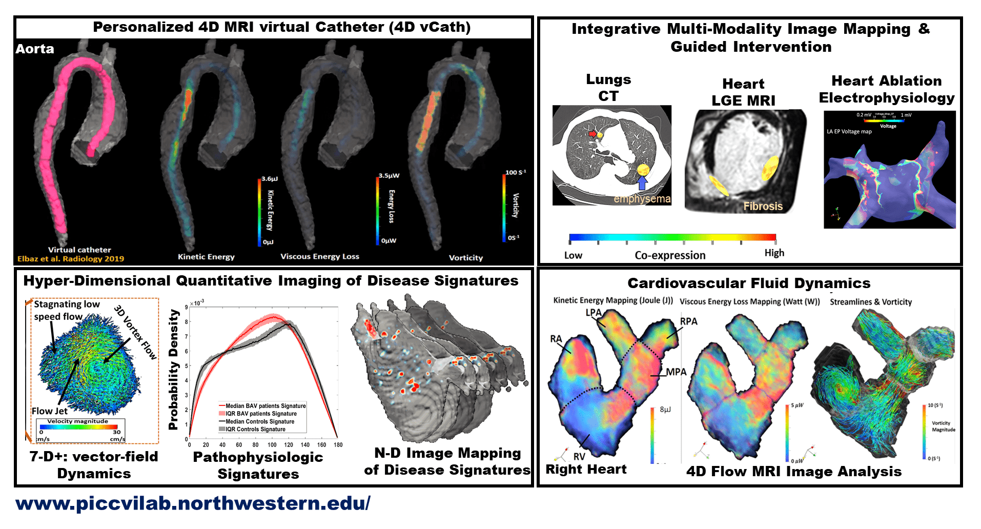
• Medical image processing • 4D MRI virtual catheter • Computational cardiovascular imaging • Quantitative MRI • Inverse imaging problems• Hyper-dimensional imaging quantification • Integrative multi-modality image mapping • Personalized imaging-guided intervention • Cardiovascular fluid dynamics • Mapping disease signatures • Translational cardiovascular image analysis
Example Research Projects
4D MRI Virtual Catheter (vCath)
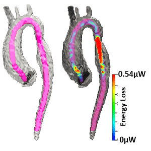
Previous in vivo and computational studies have provided evidence of the critical role of regional hemodynamic alterations on cardiovascular and neurovascular disease development. Several imaging modalities and computational tools (MRI, Doppler Echo, CFD-assisted CT, patient-specific CFD) can measure or provide noninvasive blood flow velocities but no approach is available that provide intuitive quantification or visualization of blood flow in a manner similar to the invasive Cath that is most familiar to clinicians. This invention address this unmet need via a method for generating a virtual Catheter (vCath) that uses mathematical modeling to mimic the well-established invasive catheter in probing hemodynamics but from any noninvasive modality or computational flow modeling that provide flow information.
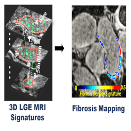
Precise Fibrosis Signatures for MRI-Guided Cardiac Diagnosis & Intervention
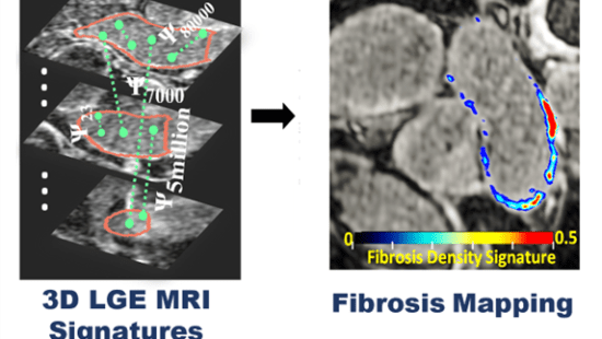
Left atrial (LA) fibrosis assessed with 3D (LGE)-MRI has shown promise in evaluating atrial myopathy, and predicts AF recurrence [1, 2]. Current methods for fibrosis quantification suffer from lack of standardization and reproducibility as they rely on different thresholds for defining fibrosis which hinders effective clinical translation of LGE-MRI. This issue can lead to the false candidacy of patients for catheter ablation and affects life expectancy in Afib patients. To address these limitations, we introduced the first threshold-free technique for objectively quantifying fibrosis burden from 3D LGE [3]. Our proposed method maps each 3D LGE MRI patient data to its signatures. Fig.1 represents stages of computing Fibrosis Signature Index (FSI) in more details which derives a unique co-association of multi-millions of 3d LGE-MRI intensities of left atrium. The computation cost of this method is also low as this method is done stochastically. Interested readers can refer to 2022 ISMRM abstract “Stochastic Fibrosis Signatures from 3D LGE: Novel Threshold-Free quantification of left atrial Fibrosis” for more detail. We also expand this project including but not limited to assess impact of segmentation, image resolution and artifact on this method. We also work on an automated segmentation error in 3D LGE MRI.
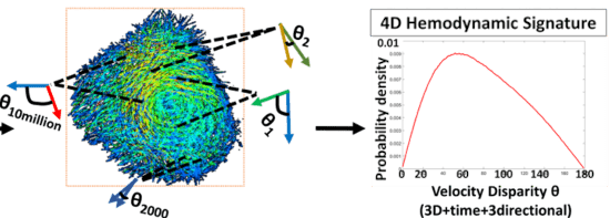
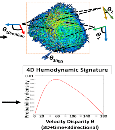
Hyper-dimensional 4D Flow MRI Signatures of Composite Blood Flow Dynamics
4D Flow provides a 3D time-resolved velocity vector field consisting of 3-directional velocity vectors in each image voxel1,2. This powerful modality can be used to visualize fluid flow throughout the heart with the potential to quantify the blood flow dynamics of cardiac diseases. To fully utilize this rich data, our lab has developed a novel 4D Flow Vector-Field Signature technique that quantifies intracardiac flow through stochastic probabilistic analysis3. By stochastically sampling paired vector disparities across cardiac volumes and time, 4D Flow Vector-Field Signatures comprehensively capture complex blood flow dynamics to create standardized profiles that are easily comparable between patients.
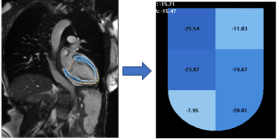
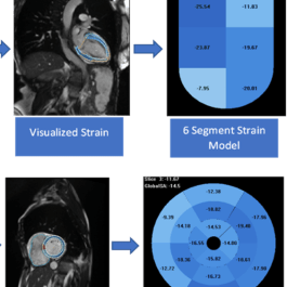
Cardiac Mechanics by MRI Strain
Left Ventricular (LV) strain is generally reported as a global measure (averaged across the entire ventricle) due to its high reproducibility. Strain can be calculated at a more regional level as well, generally based on the 16 segments of the LV outlined by the AHA, but this type of segmental strain is not used clinically due to its lower reproducibility. However, regional strain measurements have the potential of being important localized biomarkers to track cardiac disease and responses to treatment.
Example Research Projects
4D MRI Virtual Catheter (vCath)
Hyper-dimensional 4D Flow MRI Signatures of Composite Blood Flow Dynamics
Precise Fibrosis Signatures for MRI-Guided Cardiac Diagnosis & Intervention