
4D Blood Flow Dynamics in the Heart
The heart acts as a pump, driving the circulation of blood throughout the body. In many cardiac diseases, this pumping action can be impacted, leading to health complications across the body. In order to better understand the impacts of cardiac diseases on the blood flow dynamics of the heart, we use a magnetic resonance imaging (MRI) technique called 4D Flow.
4D Flow provides a 3D time-resolved velocity vector field consisting of 3-directional velocity vectors in each image voxel1,2. This powerful modality can be used to visualize fluid flow throughout the heart with the potential to quantify the blood flow dynamics of cardiac diseases. To fully utilize this rich data, our lab has developed a novel 4D Flow Vector-Field Signature technique that quantifies intracardiac flow through stochastic probabilistic analysis3. By stochastically sampling paired vector disparities across cardiac volumes and time, 4D Flow Vector-Field Signatures comprehensively capture complex blood flow dynamics to create standardized profiles that are easily comparable between patients.
References:
[1] Markl M, Frydrychowicz A, Kozerke S, Hope M, Wieben O. 4D flow MRI. Journal of Magnetic Resonance Imaging. 2012;36(5):1015-1036.
[2] Dyverfeldt P, Bissell M, Barker AJ, et al. 4D flow cardiovascular magnetic resonance consensus statement. Journal of Cardiovascular Magnetic Resonance. 2015;17(1):72. doi:10.1186/s12968-015-0174-5
[3] Elbaz MSM, Malaisrie C, McCarthy P, Markl M. Stochastic 4D Flow Vector-Field Signatures: A New Approach for Comprehensive 4D Flow MRI Quantification. In: de Bruijne M, Cattin PC, Cotin S, et al., eds. Medical Image Computing and Computer Assisted Intervention – MICCAI 2021. Lecture Notes in Computer Science. Springer International Publishing; 2021:215-224.
Aorta
In bicuspid aortic valve disease (BAV), the valve leading from the left ventricle to the aorta has an altered geometry. This can lead to backflow into the left ventricle when the heart pumps (regurgitation) and a narrowing of the aorta (stenosis). 4D flow MRI can provide insight into the effects of BAV on blood flow dynamics in the aorta, potentially providing more information about the clinical severity of the disease. The 4D vector-field signature technique was developed in the aorta in order to capture both local and global flow dynamic changes that are associated with BAV. Applications of this quantification method may lead to a better understanding of the risk factors for the severe and potentially life-threatening consequences of BAV such as aortic dissection.
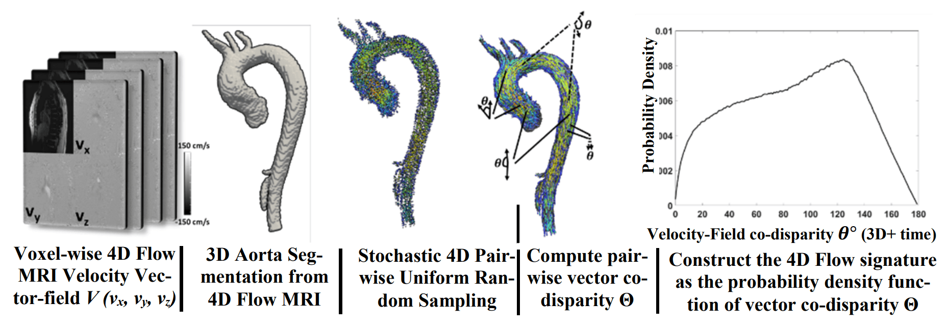
Figure: 4D Flow MRI image (left) provides an anatomical magnitude image, as well as quantitative images of each velocity direction (x,y,z). These can be combined to create a 3-directional, time-resolved velocity vector-field of anatomical structures such as the aorta. The 4D Flow Vector-Field Signature (right) is created by pair-wise probabilistic sampling throughout the velocity vector-field to create standardized profiles of blood flow dynamics.
Publications:
[1] Elbaz MSM, Malaisrie C, McCarthy P, Markl M. Stochastic 4D Flow Vector-Field Signatures: A New Approach for Comprehensive 4D Flow MRI Quantification. In: de Bruijne M, Cattin PC, Cotin S, et al., eds. Medical Image Computing and Computer Assisted Intervention – MICCAI 2021. Lecture Notes in Computer Science. Springer International Publishing; 2021:215-224.
[2] Elbaz MSM, Scott MB, Barker AJ, Ryan Avery McCarthy P, Malaisrie C, , Bonow RO, Carr J and Markl M. 4D Flow Hemodynamic Signatures: A novel technique for assessing hemodynamics in aortic valve disease – evaluation in 418 patients and controls disease. ”, Society for Cardiovascular Magnetic Resonance (SCMR) 23rd Scientific Meeting, Orlando, Florida, February 12-15, 2020
Patents
Elbaz, Mohammed SM, et al. “Co-Expression Signatures Method for Quantification of Physiological and Structural Data.” U.S. Patent Application No. 17/004,779.
Left Atrium
Atrial fibrillation (AF) is a heart arrhythmia experienced by over 30 million people worldwide. Disruptions in the electrical rhythms of the heart can lead to severe impacts on blood flow dynamics, such as causing blood to stagnate and clot in the left atrial chamber1,2,3. When clots are pumped through the body and land in the brain, this can cause a stroke. AF patients are at an increased risk of stroke, but stroke risk assessment does not take the individual blood flow dynamics of a patient’s heart into account, potentially missing vital clinical information.
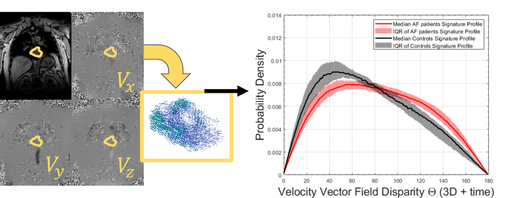
Left atrial blood flow quantification in the presence of atrial fibrillation (AF) is an important clinical question but is complex because left atrial flow can consist of multiple interacting fluid dynamics components such as vortices, regurgitant jets, and stagnations4,5. A comprehensive quantification technique is required. To address current shortcomings, we have extended our 4D Flow Vector-Field Signature technique for use in the left atrium, capturing alterations in 4D hemodynamics associated with AF6. This approach can be leveraged to better characterize the pathophysiology of AF flow7, and to contribute to a long-term goal of understanding the etiology of AF-induced stroke risk.
Figure: 4D Flow MRI image (left) provides an anatomical magnitude image, as well as quantitative images of each velocity direction (x,y,z). These can be combined to create a 3-directional, time-resolved velocity vector-field of anatomical structures such as the left atrium. The 4D Flow Vector-Field Signature (right) is created by probabilistic sampling throughout the velocity vector-field to create standardized profiles that capture blood flow dynamics alterations due to cardiac diseases such as atrial fibrillation (red).
Patents
Elbaz, Mohammed SM, et al. “Co-Expression Signatures Method for Quantification of Physiological and Structural Data.” U.S. Patent Application No. 17/004,779.
Publications:
[1] Nallamothu T, DiCarlo AL, Lee DC, Kim D, Arora R, Markl M, Greenland P, Passman RS, Elbaz MSM. Novel Stochastic 4D Flow Signatures of time-resolved 3D left atrial flow-field alterations in atrial fibrillation. The annual conference of the International Society for Magnetic Resonance in Medicine (ISMRM), 15-20 May 2021.
[2] Nallamothu T, Pradella M, Lee DC, Kim D, Markl M, Greenland P, Passman RS, Elbaz MSM. Novel atrial 4D Flow Hemodynamic Signature Index (HSI) associates inversely with stroke volume and directly with LA size independent of age, gender, and CHA₂DS₂-VASc score in atrial fibrillation patients. Society for Cardiovascular Magnetic Resonance (SCMR) 25th annual meeting, February 2-5, 2022.
Awards:
- SCMR 2022 Early Career Award Finalist.
- SCMR 2022 Travel Award
- ISMRM 2021 Best Cardiac Trainee Abstract.
- ISMRM 2021 Magna Cum Laude.
- ISMRM2021 Trainee Stipend Award.
References:
[1] Goldman ME, Pearce LA, Hart RG, et al. Pathophysiologic Correlates of Thromboembolism in Nonvalvular Atrial Fibrillation: I. Reduced Flow Velocity in the Left Atrial Appendage (The Stroke Prevention in Atrial Fibrillation [SPAF-III] Study). Journal of the American Society of Echocardiography. 1999;12(12):1080-1087.
[2] Handke M, Harloff A, Hetzel A, Olschewski M, Bode C, Geibel A. Left Atrial Appendage Flow Velocity as a Quantitative Surrogate Parameter for Thromboembolic Risk: Determinants and Relationship to Spontaneous Echocontrast and Thrombus Formation–A Transesophageal Echocardiographic Study in 500 Patients with Cerebral Ischemia. Journal of the American Society of Echocardiography. 2005;18(12):1366-1372.
[3] Pollick C, Taylor D. Assessment of left atrial appendage function by transesophageal echocardiography. Implications for the development of thrombus. Circulation. 1991;84(1):223-231.
[4] Föll D, Taeger S, Bode C, Jung B, Markl M. Age, gender, blood pressure, and ventricular geometry influence normal 3D blood flow characteristics in the left heart. European Heart Journal – Cardiovascular Imaging. 2013;14(4):366-373.
[5] Fyrenius A, Wigström L, Ebbers T, Karlsson M, Engvall J, Bolger AF. Three dimensional flow in the human left atrium. Heart. 2001;86(4):448-455.
[6] Nallamothu T, DiCarlo AL, Lee DC, Kim D, Arora R, Markl M, Greenland P, Passman RS, Elbaz MSM. Novel Stochastic 4D Flow Signatures of time-resolved 3D left atrial flow-field alterations in atrial fibrillation. The annual conference of the International Society for Magnetic Resonance in Medicine (ISMRM), 15-20 May 2021.
[7] Nallamothu T, Pradella M, Lee DC, Kim D, Markl M, Greenland P, Passman RS, Elbaz MSM. Novel atrial 4D Flow Hemodynamic Signature Index (HSI) associates inversely with stroke volume and directly with LA size independent of age, gender, and CHA₂DS₂-VASc score in atrial fibrillation patients. Society for Cardiovascular Magnetic Resonance (SCMR) 25th annual meeting, February 2-5, 2022.
4D MRI Error Correction
4D flow MRI images are created by using fast-switching magnetic field gradients to capture the velocity of moving tissue, such as blood. These quickly changing gradients can cause eddy currents in the MRI hardware, leading to inaccuracies in the velocity measurements that vary across the space in the scanner1,2.
Importantly, these errors can be different in each velocity encoding direction, leading to spatially varying inaccuracies in both the magnitude and direction of velocity vectors throughout the acquired vector field. Many methods exist to current these errors, but the optimizations of such methods have significant shortcomings (time-consuming, not generalizable). We have developed a new approach for optimizing eddy current corrections based on our 4D Flow Vector-Field Signature technique3. Eddy current-induced heterogeneity in the velocity vector field is captured in the shape of 4D Flow Vector-Field Signature profiles, but this metric is also stable and robust to noise, allowing for convergence when errors due to eddy currents are minimized. By utilizing the inherent stability of the 4D Flow Vector-Field Signatures to correct the underlying velocity vector field data, our approach is self-calibrating and generalizable to different scan protocols.

Figure: 4D Flow MRI image (left) provides an anatomical magnitude image, as well as quantitative images of each velocity direction (x,y,z). These can be combined to create a 3-directional, time-resolved velocity vector-field of anatomical structures. Eddy current errors due to fast-switching magnetic field gradients inherent to 4D Flow can lead to offsets in each velocity direction () leading to the corruption in the magnitude and direction of measured velocity vectors across the velocity vector field.
Publications:
[1] Nallamothu T, Berhane H, Ma L, Baraboo J, Lee DC, Kim D, Greenland P, Markl M, Passman RS, Elbaz MSM. Self-calibrating method to simultaneously optimize eddy current correction and detect wraparound artifacts using a novel stochastic 4D flow-field disparity signature technique. Society for Magnetic Resonance Angiography 33rd Annual International Conference, 9-12 September 2021.
[2] Nallamothu T, Berhane H, Ma L, Baraboo J, Lee DC, Kim D, Greenland P, Markl M, Passman R, Elbaz MSM. Novel Self-calibrating 4D Flow-Field Signature Technique to Simultaneously Optimize Eddy Current Corrections and Detect Wraparound Errors. The annual conference of the International Society for Magnetic Resonance in Medicine (ISMRM), 7-12 May 2022.
Awards:
ISMRM 2022 Best Flow and Motion Poster Finalist.
ISMRM 2022 Educational Stipend.
Patents
Elbaz, Mohammed SM, et al. “Co-Expression Signatures Method for Quantification of Physiological and Structural Data.” U.S. Patent Application No. 17/004,779.
References:
[1] Markl M, Frydrychowicz A, Kozerke S, Hope M, Wieben O. 4D flow MRI. Journal of Magnetic Resonance Imaging. 2012;36(5):1015-1036.
[2] Lorenz R, Bock J, Snyder J, Korvink JG, Jung BA, Markl M. Influence of eddy current, Maxwell and gradient field corrections on 3D flow visualization of 3D CINE PC-MRI data. Magnetic Resonance in Medicine. 2014;72(1):33-40
[3] Nallamothu T, Pradella M, Lee DC, Kim D, Markl M, Greenland P, Passman RS, Elbaz MSM. Novel atrial 4D Flow Hemodynamic Signature Index (HSI) associates inversely with stroke volume and directly with LA size independent of age, gender, and CHA₂DS₂-VASc score in atrial fibrillation patients. Society for Cardiovascular Magnetic Resonance (SCMR) 25th annual meeting, February 2-5, 2022.