
Left atrial (LA) fibrosis assessed with 3D (LGE)-MRI has shown promise in evaluating atrial myopathy, and predicts AF recurrence [1, 2]. Current methods for fibrosis quantification suffer from lack of standardization and reproducibility as they rely on different thresholds for defining fibrosis which hinders effective clinical translation of LGE-MRI. This issue can lead to the false candidacy of patients for catheter ablation and affects life expectancy in Afib patients. To address these limitations, we introduced the first threshold-free technique for objectively quantifying fibrosis burden from 3D LGE [3]. Our proposed method maps each 3D LGE MRI patient data to its signatures. Fig.1 represents stages of computing Fibrosis Signature Index (FSI) in more details which derives a unique co-association of multi-millions of 3d LGE-MRI intensities of left atrium. The computation cost of this method is also low as this method is done stochastically. Interested readers can refer to 2022 ISMRM abstract “Stochastic Fibrosis Signatures from 3D LGE: Novel Threshold-Free quantification of left atrial Fibrosis” for more detail. We also expand this project including but not limited to assess impact of segmentation, image resolution and artifact on this method. We also work on an automated segmentation error in 3D LGE MRI.
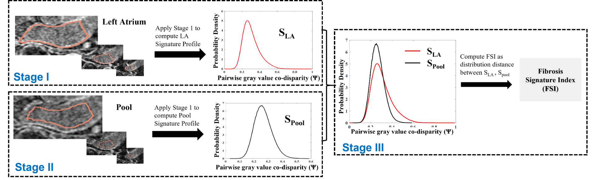
Fig.1 Illustration of stage I, II and III of the stochastic fibrosis signatures technique from 3D LGE MRI. Stage I computes a unique profile of co-assosiation of LGE-MRI intensities over entire LA which is derived stochastically, We call it LA signature profile.
[1] Dzeshka MS, Lip GY, Snezhitskiy V, and Shantsila E. “Cardiac Fibrosis in Patients With Atrial Fibrillation: Mechanisms and Clinical Implications”. J Am Coll Cardiol. 2015;66:943-59.
[2] Marrouche NF, Wilber D, Hindricks G, Jais P, Akoum N, Marchlinski F, Kholmovski E, Burgon N, Hu N, Mont L, Deneke T, Duytschaever M, Neumann T, Mansour M, Mahnkopf C, Herweg B, Daoud E, Wissner E, Bansmann P and Brachmann J. Association of atrial tissue fibrosis identified by delayed enhancement MRI and atrial fibrillation catheter ablation: the DECAAF study. JAMA. 2014; 311:498-506.
[3] Mehrnia M, Kholmovski E, Passman R, Katsaggelos A, Nazarian S, Kim D, and Mohammed Elbaz, “Stochastic Fibrosis Signatures from 3D LGE: Novel Threshold-Free quantification of left atrial Fibrosis”, ISMRM 2022.
Punlications
- Mehri Mehrnia, Eugene Kholmovski, Daniel Kim, Rod Passman, Mohammed S. M. Elbaz. “Novel Threshold-free Fibrosis Signature Index (FSI) for quantification of left atrial fibrosis and scar burden from 3D LGE”. In Proceedings: Society for Cardiovascular Magnetic Resonance (SCMR) 26th annual meeting, January 25-28, 2023 (Accepted)
- Mehrnia M, Kholmovski E, Passman R, Katsaggelos A, Nazarian S, Kim D and Elbaz MS. Stochastic Fibrosis Signatures from 3D LGE: Novel Threshold-Free quantification of left atrial fibrosis. In Proceedings: International Society of Magnetic Resonance in Medicine (ISMRM) 31st Annual Meeting. 7-12 May, 2022.
Awards
- ISMRM 2022 Magna Cum Laude Award for the work
- ISMRM 2022 Stipend Award
Patents
Elbaz, Mohammed SM, et al. “Co-Expression Signatures Method for Quantification of Physiological and Structural Data.” U.S. Patent Application No. 17/004,779.